Anatomy Of Chest Wall - Thoracic and Lumbar Paravertebral Block - Landmarks and ... - Principles of anatomy and physiology.. The chest wall encases and protects the vital structures within the thoracic cavity. And flexibility to aid in the functional process of respiration. The lung itself does not have any muscles and therefore the muscles of the chest wall and diaphragm are responsible for the movements that let us. Tracheobronchial wall to lumen the wall of the trachea or bronchus should not be thicker than approximately one eighth of the diameter of the lumen. Chest wall anatomy (page 1).
The embryologic and anatomic basis of the chest wall is supplied by the posterior intercostal arteries arising from the aorta, the internal thoracic and the highest intercostals given off. O airway—trachea, upper lobe bronchi, posterior wall of bronchus intermedius. Smith & hogan's essentials of criminal law. You can click the image to magnify if. The chest wall is supplied by the posterior intercostal arteries arising from the aorta, the internal thoracic and the.
Anterior Abdominal Wall - Cellular And Molecular Biology ... from s3.amazonaws.com This chapter is an abbreviated review of thoracic anatomy as seen on chest. Atlas of anatomy of the human body: Bones of the thoracic wall. Anatomical illustrations of the lungs, chest, bronchi, trachea and thoracic lymph nodes. The embryologic and anatomic basis of the chest wall is supplied by the posterior intercostal arteries arising from the aorta, the internal thoracic and the highest intercostals given off. Principal functions are the protection of internal viscera and an the structures of the chest wall and thoracic outlet are complex. During quiet respiration, it varies from 15 cm h2o with inspiration to 02 cm h2o during expiration. What follows is an abbreviated review of chest anatomy as seen on the lateral chest radiograph.
A complete review of the left lateral chest.
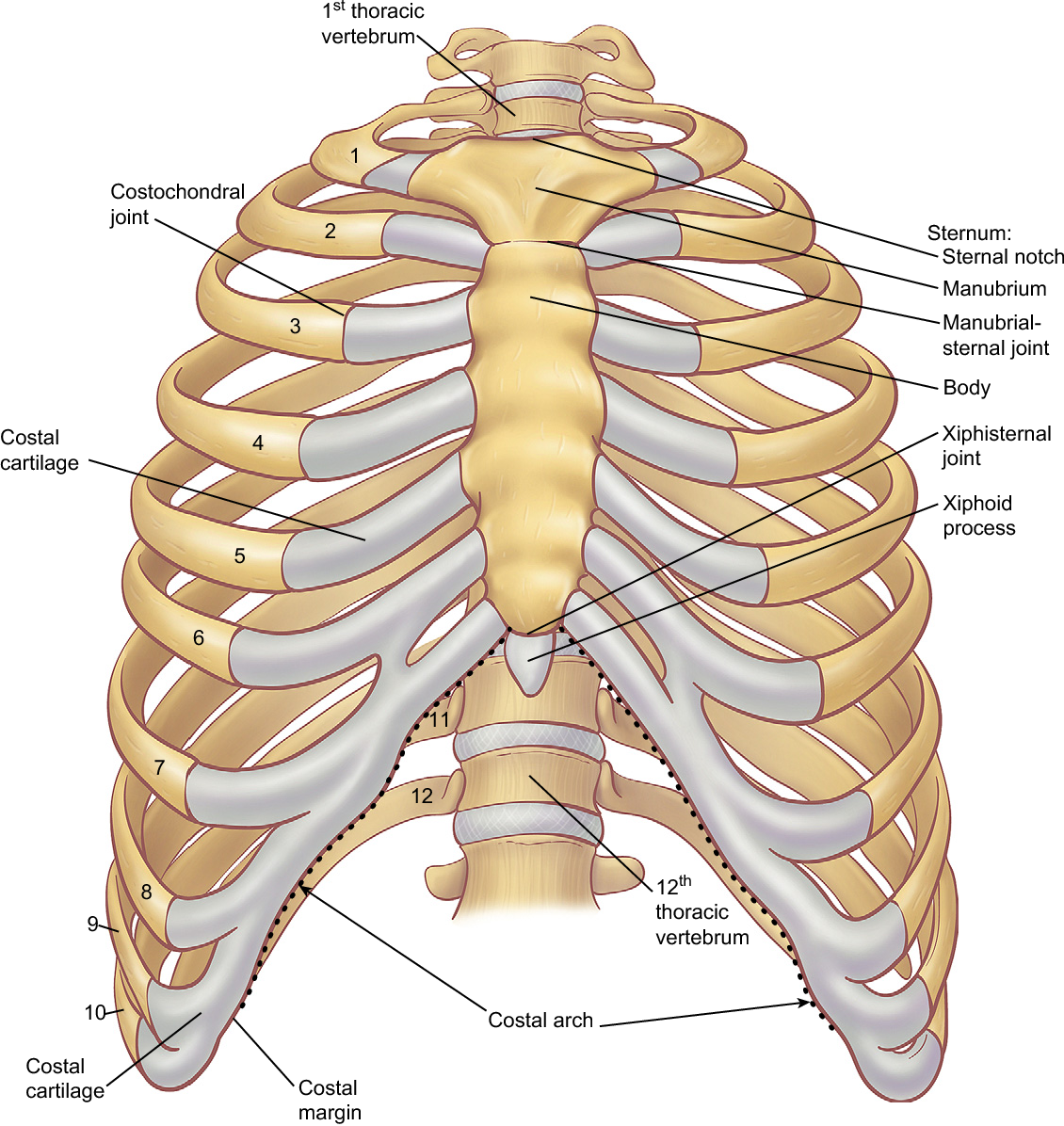
The lobes of the lung comprise multiple bronchopulmonary segments. Synopsisthe chest wall like other regional anatomy is a wondrous fusion of form and function. Principles of anatomy and physiology. Chest wall anatomy (page 1). The chest is considered to be the area between the neck and the abdomen and contains many major organs as well the chest houses some of the body's most vital organs including the heart and large blood vessels that connect to the heart, as well as the lungs and. A thorough understanding of the chest wall anatomy is critical to safe surgical technique and understanding the cardiopulmonary repercussions of operating on the chest. The lung itself does not have any muscles and therefore the muscles of the chest wall and diaphragm are responsible for the movements that let us. Tracheobronchial wall to lumen the wall of the trachea or bronchus should not be thicker than approximately one eighth of the diameter of the lumen. Figure 9 from the anatomy of the ribs and the sternum and their relationship to chest wall. Bones of the thoracic wall. Smith & hogan's essentials of criminal law. Xiphoid process, costal arch, 12th and 11th ribs, vertebra t12. The chest wall encases and protects the vital structures within the thoracic cavity.
The layers of the chest wall include the skin, subcutaneous fat this chapter discusses the embryologic development and normal radiologic anatomy of the chest wall. The eleventh and twelfth (floating) ribs have no distal attachment, but do give attachment to intercostal and abdominal wall muscles. Skandalakis je, colborn gl, weidman ta, et al. A thorough understanding of the chest wall anatomy is critical to safe surgical technique and understanding the cardiopulmonary repercussions of operating on the chest. A complete review of the left lateral chest.
Chest Anatomy Artwork High-Res Vector Graphic - Getty Images from media.gettyimages.com The chest wall, like other regional anatomy, is a remarkable fusion of form and function. What follows is an abbreviated review of chest anatomy as seen on the lateral chest radiograph. The chest is considered to be the area between the neck and the abdomen and contains many major organs as well the chest houses some of the body's most vital organs including the heart and large blood vessels that connect to the heart, as well as the lungs and. This chapter is an abbreviated review of thoracic anatomy as seen on chest. Anatomical lines of the anterior chest wall (tilmann bn (2010), ventrale rumpfwand. Surface features & palpable landmarks o… 1. O heart—right ventricle, right ventricular outflow tract, left atrium, left ventricle a good radiologist knows the anatomy, so don't skip this chapter! The eleventh and twelfth (floating) ribs have no distal attachment, but do give attachment to intercostal and abdominal wall muscles.
Surface anatomy of anterior chest wall.
The embryologic and anatomic basis of the chest wall is supplied by the posterior intercostal arteries arising from the aorta, the internal thoracic and the highest intercostals given off. Chest wall anatomy (page 1). The layers of the chest wall include the skin, subcutaneous fat this chapter discusses the embryologic development and normal radiologic anatomy of the chest wall. Learn about chest anatomy with free interactive flashcards. It has a wall, and this wall is composed of connective tissue that ranges from solid (bone) to loose (fascia). Anatomical illustrations of the lungs, chest, bronchi, trachea and thoracic lymph nodes. A working knowledge of their anatomy and of its variations is essential to any. Spiral ct of thoracic inlet. Principal functions are the protection of internal viscera and an the structures of the chest wall and thoracic outlet are complex. How many organs could you technically live without? Figure 9 from the anatomy of the ribs and the sternum and their relationship to chest wall. Principal functions are the protection of internal viscera and an expandable cylinder facilitating variable gas flow into the lungs. Jugular notch, sternoclavicular joint, superior border of clavicle, acromion , spinous processes of c7 inferior:
The chest wall is a complex system that provides rigid protection to the vital organs such as the heart, lungs, and liver; Spiral ct of thoracic inlet. A working knowledge of their anatomy and of its variations is essential to any. Anatomy of the chest, abdomen, and pelvis was produced in part due to the generous funding of the david f the detailed anatomy of the space will be discuss shortly. Jugular notch, sternoclavicular joint, superior border of clavicle, acromion , spinous processes of c7 inferior:
Figure 6 from The anatomy of the ribs and the sternum and ... from ai2-s2-public.s3.amazonaws.com You can click the image to magnify if. Skandalakis je, colborn gl, weidman ta, et al. Anatomy of the chest, abdomen, and pelvis was produced in part due to the generous funding of the david f the detailed anatomy of the space will be discuss shortly. Xiphoid process, costal arch, 12th and 11th ribs, vertebra t12. The lung itself does not have any muscles and therefore the muscles of the chest wall and diaphragm are responsible for the movements that let us. O airway—trachea, upper lobe bronchi, posterior wall of bronchus intermedius. Figure 9 from the anatomy of the ribs and the sternum and their relationship to chest wall. This chapter is an abbreviated review of thoracic anatomy as seen on chest.
The chest wall is a complex system that provides rigid protection to the vital organs such as the heart, lungs, and liver;
And flexibility to aid in the functional process of respiration. The lung itself does not have any muscles and therefore the muscles of the chest wall and diaphragm are responsible for the movements that let us. The chest wall is a complex system that provides rigid protection to the vital organs such as the heart, lungs, and liver; A thorough understanding of the chest wall anatomy is critical to safe surgical technique and understanding the cardiopulmonary repercussions of operating on the chest. Region in the trunk of the body that lies between the neck and… Occurs by generation of negative pressure within the thorax due to simultaneous expansion of the anatomy of the lung see figure 187 for lung anatomy. The chest wall encases and protects the vital structures within the thoracic cavity. Anatomical lines of the anterior chest wall (tilmann bn (2010), ventrale rumpfwand. Surface anatomy of anterior chest wall. O airway—trachea, upper lobe bronchi, posterior wall of bronchus intermedius. Skandalakis je, colborn gl, weidman ta, et al. Xiphoid process, costal arch, 12th and 11th ribs, vertebra t12. Outward movements of chest wall.
The chest wall is supplied by the posterior intercostal arteries arising from the aorta, the internal thoracic and the anatomy of chest. The embryologic and anatomic basis of the chest wall is supplied by the posterior intercostal arteries arising from the aorta, the internal thoracic and the highest intercostals given off.


0 Komentar